Advanced Quantitative Fluorescence Microscopy to Probe the Molecular Dynamics of Viral Entry, Science Lab
Por um escritor misterioso
Descrição
Viral entry into the host cell requires the coordination of many cellular and viral proteins in a precise order. Modern microscopy techniques are now allowing researchers to investigate these interactions with higher spatiotemporal resolution than ever before. Here we present two examples from the field of HIV research that make use of an innovative quantitative imaging approach as well as cutting edge fluorescence lifetime-based confocal microscopy methods to gain novel insights into how HIV fuses to cell membranes and enters the cell.
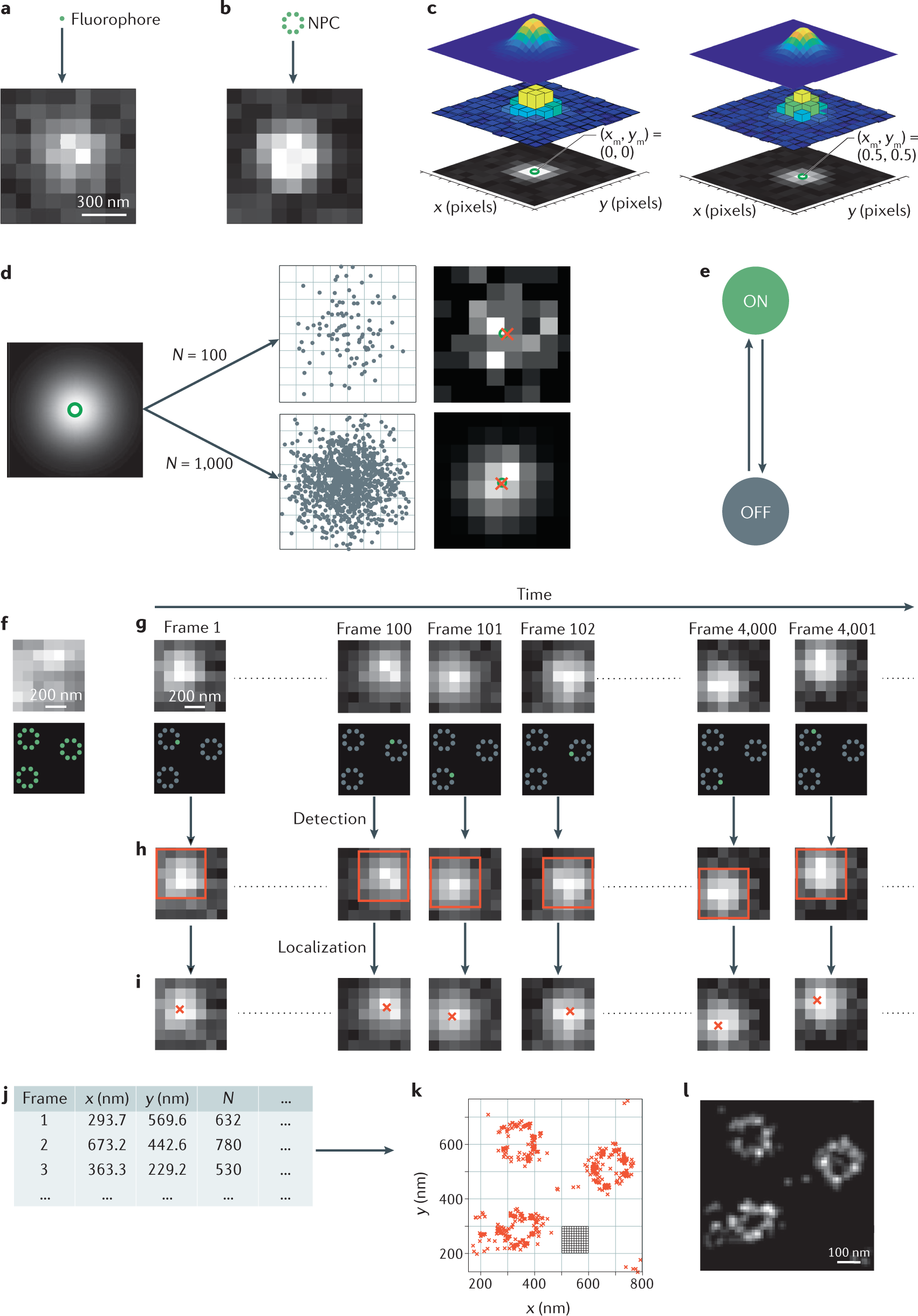
Single-molecule localization microscopy

Labeling of virus components for advanced, quantitative imaging analyses - Sakin - 2016 - FEBS Letters - Wiley Online Library
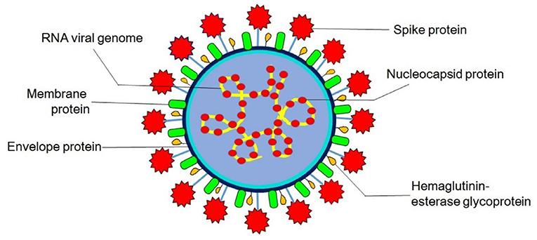
Frontiers Evolution, Interspecies Transmission, and Zoonotic Significance of Animal Coronaviruses
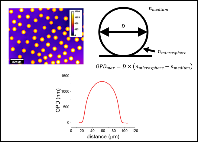
Measurement Assurance for Quantitative Cell Imaging by Optical Microscopy

Mucins Inhibit Coronavirus Infection in a Glycan-Dependent Manner

Macrophage Sult2b1 promotes pathological neovascularization in age-related macular degeneration

A comparison of conventional and advanced electroanalytical methods to detect SARS-CoV-2 virus: A concise review - ScienceDirect

A chimeric virus-based probe unambiguously detects live circulating tumor cells with high specificity and sensitivity - ScienceDirect
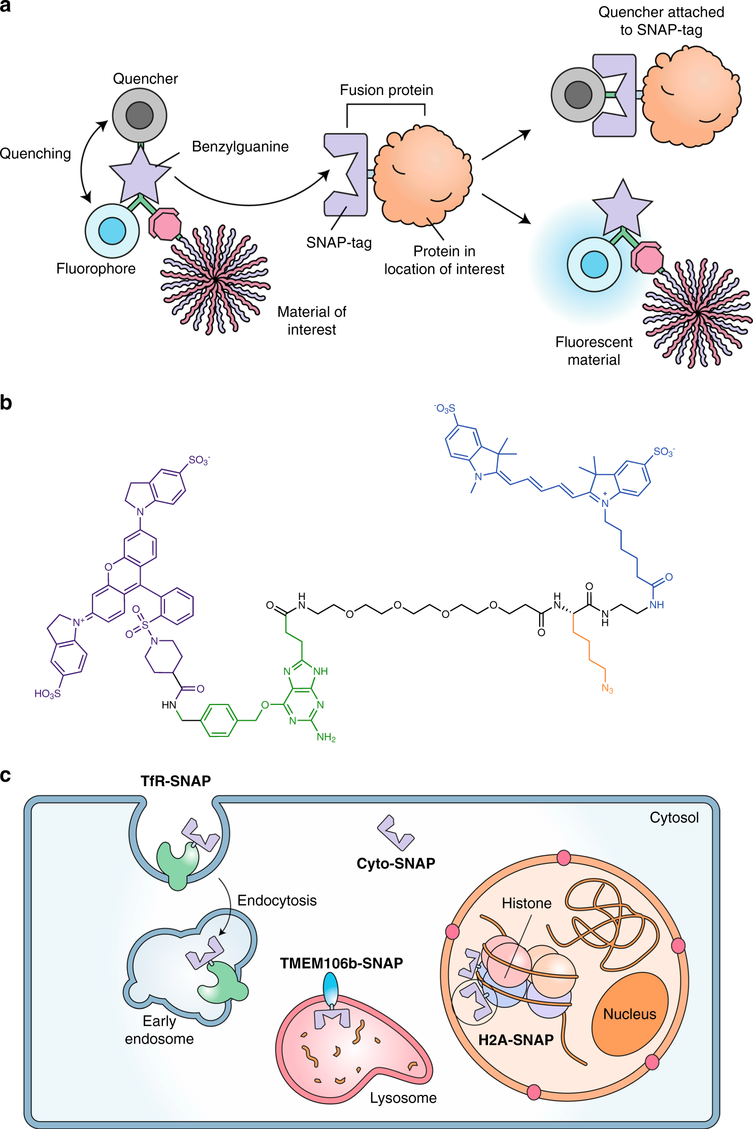
A molecular sensor to quantify the localization of proteins, DNA and nanoparticles in cells

Labeling of virus components for advanced, quantitative imaging analyses - Sakin - 2016 - FEBS Letters - Wiley Online Library

Renal-Clearable Molecular Probe for Near-Infrared Fluorescence Imaging and Urinalysis of SARS-CoV-2
de
por adulto (o preço varia de acordo com o tamanho do grupo)